Lower Body Skeletal Anatomy : The Bones Of The Lower Body Stock Image F001 7770 Science Photo Library - In turn, the pelvic girdle consists of two hip bones and the sacrum, interconnected at the pubic symphysis and sacroiliac joints.
byAdmin-
0
Lower Body Skeletal Anatomy : The Bones Of The Lower Body Stock Image F001 7770 Science Photo Library - In turn, the pelvic girdle consists of two hip bones and the sacrum, interconnected at the pubic symphysis and sacroiliac joints.. This curve, called lordosis, helps to: The back muscles are skeletal muscles. The musculoskeletal system (locomotor system) is a human body system that provides our body with movement, stability, shape, and support.it is subdivided into two broad systems: Anatomy of the skeletal system. The structural framework of the hip region is provided by the pelvis, a structure composed of the pelvic girdle and the coccyx.
1 your spine in this region has a natural inward curve. The heart and lungs are located within the thoracic cavity, and the vertebral column provides structure and protection for the spinal cord. It consists of the skull, vertebral column (including the sacrum and coccyx), and the thoracic cage, formed by the ribs and sternum. The axial skeleton supports the head, neck, back, and chest and thus forms the vertical axis of the body. Hence, it is not surprising that the bones forming the.
Lower Body Bones High Resolution Stock Photography And Images Alamy from c8.alamy.com Here is a depiction of the skeletal frame with the lower. Teachme anatomy part of the teachme series the medical information on this site is provided as an information resource only, and is not to be used or relied on for any diagnostic or treatment purposes. *the origin, insertion, and belly.* a muscle's origin is where a tendon attaches it to the *less* movable bone. Discussed in this article as part of the axial skeleton is a third subdivision, the visceral, comprising the lower jaw, some elements of the upper jaw, and the branchial arches, including the hyoid bone. The thigh is that portion of the lower limb located between the hip joint and knee joint. Skeletal_anatomy_of_lower_limb 3/5 skeletal anatomy of lower limb netter correlative imaging: The sacrum has a triangular shape, with its peak facing downward. 1 your spine in this region has a natural inward curve.
Distal to the ankle is the foot.
Your lower back (lumbar spine) is the anatomic region between your lowest rib and the upper part of the buttock. The pelvic skeleton (or pelvic girdle) consists of two hip bones and the sacrum. The action refers to the action of each muscle from the standard anatomical position. The axial skeleton supports the head, neck, back, and chest and thus forms the vertical axis of the body. The iliopsoas, an anterior muscle, flexes the thigh. *the origin, insertion, and belly.* a muscle's origin is where a tendon attaches it to the *less* movable bone. Its lower part resembles a bagel. Here is a depiction of the skeletal frame with the lower. Start studying lower limb skeletal anatomy. Anatomical directional terms 12 photos of the anatomical directional terms anatomical directional terms powerpoint, anatomical orientation and directional terms, anatomical orientation and directional terms that have the same meaning, directional terms related to anatomical position, list of anatomical directional terms. Muscular system, which includes all types of muscles in the body.skeletal muscles, in particular, are the ones that act on the body joints to produce movements. Tibia and fibula in anatomical position with parts labeled. It consists of the bones that make up the arms and legs, as well as the bones that attach them to the.
Learn vocabulary, terms, and more with flashcards, games, and other study tools. Hence, it is not surprising that the bones forming the. *the origin, insertion, and belly.* a muscle's origin is where a tendon attaches it to the *less* movable bone. The lower limbs carry the total body weight when we are erect; The skeleton is subdivided into two divisions:
Jesse Young S Human Anatomy Drawing Of Skeletal Structure Of The Lower Body Circa 2005 Art Print By Jesseyoung Society6 from ctl.s6img.com Anatomical directional terms 12 photos of the anatomical directional terms anatomical directional terms powerpoint, anatomical orientation and directional terms, anatomical orientation and directional terms that have the same meaning, directional terms related to anatomical position, list of anatomical directional terms. The skeleton acts as a scaffold by providing support and protection for the soft tissues that make up the rest of the body. The heart and lungs are located within the thoracic cavity, and the vertebral column provides structure and protection for the spinal cord. It consists of skull, vertebral column, and thoracic cage. Start studying lower limb skeletal anatomy. The brain is surrounded by bones that form part of the skull. The vertebral column of the lower back includes the five lumbar vertebrae, the sacrum, and the coccyx. The skeleton protects vital organs.
Learn vocabulary, terms, and more with flashcards, games, and other study tools.
Anatomy of the skeletal system. The skeleton protects vital organs. The muscles in the medial compartment adduct the thigh. The musculoskeletal system (locomotor system) is a human body system that provides our body with movement, stability, shape, and support.it is subdivided into two broad systems: *the origin, insertion, and belly.* a muscle's origin is where a tendon attaches it to the *less* movable bone. Its lower part resembles a bagel. The lower back is really composed of three areas of the body: 1 your spine in this region has a natural inward curve. The appendicular skeleton is made up of all bones of the upper and lower limbs. Hence, it is not surprising that the bones forming the. The iliopsoas, an anterior muscle, flexes the thigh. Balance the weight of your head on top of your spine evenly distribute weights from your upper body into the lower extremities Your lower back (lumbar spine) is the anatomic region between your lowest rib and the upper part of the buttock.
The lower limbs carry the total body weight when we are erect; Skeletal_anatomy_of_lower_limb 3/5 skeletal anatomy of lower limb netter correlative imaging: The heart and lungs are located within the thoracic cavity, and the vertebral column provides structure and protection for the spinal cord. Interactions between the skeleton, muscles, and nerves move the body. Browse 222 lower back skeleton stock photos and images available, or start a new search to explore more stock photos and images.
Lower Body Anatomy Drawing Novocom Top from i2.wp.com Balance the weight of your head on top of your spine evenly distribute weights from your upper body into the lower extremities The skeleton is subdivided into two divisions: Every skeletal muscle has three main parts: They support bones, in this case, the vertebrae. The lower spine, the hips and tailbone, and the abdomen. The lower limb skeleton includes the cingulum membri inferioris (the lower limbs bones belt) and skeleton membri inferioris liberi. The skeleton of the lower limbs and the vertical spinal column is a unique evolutionary device that allowed a person to raise his head above all other living creatures on our planet. The leg is specifically the region between the knee joint and the ankle joint.
The musculoskeletal system (locomotor system) is a human body system that provides our body with movement, stability, shape, and support.it is subdivided into two broad systems:
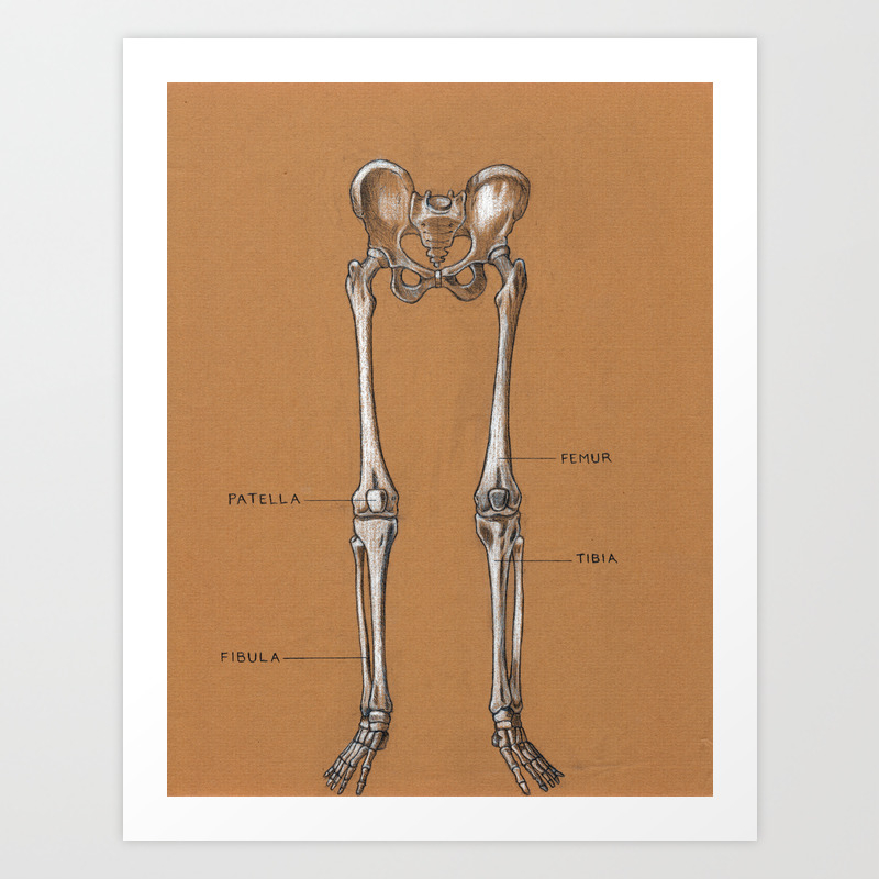
The musculoskeletal system (locomotor system) is a human body system that provides our body with movement, stability, shape, and support.it is subdivided into two broad systems: The vertebral column of the lower back includes the five lumbar vertebrae, the sacrum, and the coccyx. Lower limb bones anatomy • bones of the lower extremity interactive tutorials about the lower limb bones, lower limb bones, os coxae, femur, patella, tibia, fibula, tarsal and foot bones, featuring images, diagrams and the beautiful illustrations of getbodysmart. Our cranium (skull) protects our brain and eyes, the ribs protect our heart and lungs and our vertebrae (spine, backbones) protect our spinal cord. They support bones, in this case, the vertebrae. The skeleton acts as a scaffold by providing support and protection for the soft tissues that make up the rest of the body. Discussed in this article as part of the axial skeleton is a third subdivision, the visceral, comprising the lower jaw, some elements of the upper jaw, and the branchial arches, including the hyoid bone. The sacrum has a triangular shape, with its peak facing downward. Distal to the ankle is the foot. The lower limb contains 30 bones. The axial skeleton forms the axis of the human body. This curve, called lordosis, helps to: This bone is called the ischium.